April 26, 2024
Q&A: Dr. Ian Bookman talks colon cancer awareness, prevention, and saving lives through the Bum Run
 Learn how AI is improving our care with this interactive graphic
 Behind the Mask: Jean-Paul Michael
 We’re hiring! Explore careers at Unity Health

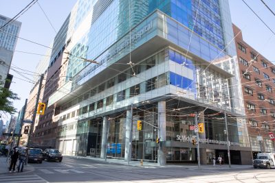
30 Bond St.,
Toronto, ON
M5B 1W8
The main entrance is located near Queen St. East and Victoria St. The Bond St. entrance is not open to the public at this time.
416-360-4000